Understand about stone disease

You may have been diagnosed with a kidney stone... Understand how this diagnosis affects you and what solutions are available to you.

This information pages cannot replace the role and relationship you have with your physician. It is essential you seek professional clinical opinion from your physician.

General information about the stone disease

General information about the stone disease

Learn more about the stone disease
Close

What is the urinary system?

References

1: Alelign T, Petros B. Kidney Stone Disease: An Update on Current Concepts. Adv Urol. 2018 Feb 4;2018:3068365.
2: D'Alessandro C, Ferraro PM, Cianchi C, Barsotti M, Gambaro G, Cupisti A. Which Diet for Calcium Stone Patients: A Real-World Approach to Preventive Care? Nutrients. 2019 May 27;11(5):1182. 
3: Patti L, Leslie SW. Acute Renal Colic. 2021 Aug 12. In: StatPearls [Internet]. Treasure Island (FL): StatPearls Publishing; 2021 Jan.
4: Xavier A, Maxwell AP. Which patients with renal colic should be referred? Practitioner. 2011 Feb;255(1737):15-7, 2. 

What is the urinary system?

It is all the organs that ensure the production and elimination of urine. It consists of two kidneys (production site), two ureters (tubes from kidney to bladder), a bladder (storage site) and a urethra (tube to urinate).

What is an urinary calculus?

It is a stone, mass of different crystals, that forms in the urinary system.

Diagram of the urinary system

Who can get urinary stone disease?

Urinary stone affects about 12% of the world population and occurs more frequently in men than in women within the age of 20-49 years.1 An increase in new cases (incidence) has been noted in the past decades in both developed and developing countries.1

Why do calculi form?2

The main reasons are diet excesses (excess calcium, sugar, salt, animal protein, etc.) and insufficient fluid intake.

What are the symptoms associated with these stones?1

When they migrate into the urinary tract, the stones may lead to the appearance of blood in the urine (hematuria), pain in the lower back (renal colic), or urinary infections.

What is an attack of renal colic?3,4

Classically, the following are signs of renal colic:

  • Sudden onset;
  • Sharp pain in the lower back (lumbar pain), on the right or left side;
  • Pain that may spread down to the testicles in men and the labia majora (vulva) in women;
  • There may be vomiting/nausea;
  • Often the patient is agitated due the intense pain and cannot find a position in which to feel more comfortable;
  • Sometimes there may be signs of irritation in the bladder (frequent desire to urinate): this generally means that the stone is at the end of the ureter at the entrance to the bladder

Why does an attack of renal colic occur?3

The stones form an obstacle to the smooth flow of urine, which accumulates above the obstacle and leads to distension of the cavities in the kidney. This overpressure then causes the attack of renal colic.

What additional examinations are required?3

In order to find the cause of the renal colic, your doctor may ask you to undergo various examinations:

  • X-ray of the abdomen (KUB) and ultrasound of the kidneys and bladder
  • Scan of the abdomen and pelvis with or without contrast injection
  • Blood sample analysis
  • An analysis of your urine to check for any infection or the presence of blood
Close
Treatment Option for stone disease

Treatment options

Learn more about the different treatment options for stone disease available. Learn more about treatment options

References

1: Patti L, Leslie SW. Acute Renal Colic. 2021 Aug 12. In: StatPearls [Internet]. Treasure Island (FL): StatPearls Publishing; 2021 Jan.
2: Xavier A, Maxwell AP. Which patients with renal colic should be referred? Practitioner. 2011 Feb;255(1737):15-7, 2.
3: Ordon M, Andonian S, Blew B, Schuler T, Chew B, Pace KT. CUA Guideline: Management of ureteral calculi. Can Urol Assoc J. 2015 Nov-Dec;9(11-12): E837–E851.
4. NHS. Treatment-Kidney stones. 2019 https://www.nhs.uk/conditions/kidney-stones/treatment
5: Alyami F, Norman R. Is an overnight stay after percutaneous nephrolithotomy safe? Arab J Urol.2012 Dec; 10(4): 367–371.
6: British Association of Urological Surgeon (BAUS). Percutaneous nephrolithotomy (keyholesurgery for kidney stones) - information about your procedure. 2020 https://www.baus.org.uk/_userfiles/pages/files/Patients/Leaflets/PCNL.pdf

How is renal colic treated?

The first and most important step, is to address the pain. In an emergency, the treatment is aimed at giving you rapid relief. The most effective medications are non-steroidal anti-inflammatory agents1,2. If these are not sufficient, more powerful painkillers derived from morphine may be used. Admission to hospital is not always necessary. The stone itself is not treated in emergency.

When is admission to hospital necessary?1,2

  • In the event of fever (temperature > 38° C or 100.4°F) associated with urinary infection.
  • In the event of persistent pain in spite of painkillers.
  • If the urine is unable to exit the body - it is called anuria and it is related to the absence of urine in the bladder.

How are calculi treated?3

  • Small stones may be eliminated spontaneously by natural means. It is useful to collect them to analyse their composition. To do this, urinate into a coffee filter or tea strainer, keep the stone(s) dry in a container and follow the instructions of your doctor. But above all, don’t throw the stone(s) away! Indeed, different kinds of stones exist and the diet recommendations will be defined according to the nature of the stone analysed.
  • If the stone is not eliminated within a reasonable length of time (a few weeks) or is still painful, your urologist may suggest you undergo surgery, the method of which will depend on the size, location and nature of the stone. In all cases, remote-controlled imaging must be performed to confirm the healing.

Different kinds of surgeries available3,4

  • Extracorporeal lithotripsy: a machine is used that creates shock waves that are directed on to the stone to break it. The fragments will then be eliminated during urination.
  • Ureteroscopy: the surgeon uses a device with a camera that is passed through the urinary natural tract to remove or destroy the stone, usually with a laser.
  • Percutaneous nephrolithotomy (or PCNL) : surgical access through the back directly into the kidney (1cm incision).
  • Open surgery via laparoscopy by incision: have become exceptional. The indications are associated with very large stones stuck in the urinary tract when above techniques are not feasible.

What is ureteroscopy?4

This technique makes it possible to reach the stone through the urinary natural tract. This is done by entering a small tube fitted with a camera, called an ureteroscope, into your urethra up to the ureter and renal cavities. Your doctor will then be able to either remove an entire stone (in 1 piece) or to fragment it using various means. Hospitalisation can last a single day in an outpatient setting to several days. This depends on the stones, anatomical difficulties and the condition of the patient. Ureteroscopy is most often carried out under a general anaesthetic. At the end of the procedure it may be decided that a small tube (a single J or double J stent) is left inside your ureter for few days.

Stones removed or fragmented using a ureteroscope (camera)

What is percutaneous nephrolithotomy?

This technique consists of puncturing the kidney through the back under general anaesthetic in order to create a direct tract into the kidney. According to the indication, patients may lie flat on their stomach or on their back, so that a camera with an instrument (this is called a nephroscope) can be positioned through the tract, enabling the stones to be visualised, fragmented and extracted. At the end of the procedure a drain is often left in place through the skin (nephrostomy). Hospitalisation for this type of procedure lasts 2 to 5 days on average.5,6

Close
JJ stent placement for stone disease

Quality of life with a JJ stent

Learn what is a double J stent used for, the side effects and how long a stent stays in. Learn more about JJ stent placement

References

1: Wiseman O, Ventimiglia E, Doizi S, Kleinclauss F, Letendre J, Cloutier J, Traxer O. Effects of Silicone Hydrocoated Double Loop Ureteral Stent on Symptoms and Quality of Life in Patients Undergoing Flexible Ureteroscopy for Kidney Stone: A Randomized Multicentre Clinical Study. J Urol. 2020 Oct;204(4):769-777.
2: Barghouthy Y, Wiseman O, Ventimiglia E, Letendre J, Cloutier J, Daudon M, Kleinclauss F, Doizi S, Corrales M, Traxer O. Silicone-hydrocoated ureteral stents encrustation and biofilm formation after 3-week dwell time: results of a prospective randomized multicenter clinical study. World J Urol. 2021 Mar 10.
3: Hofmann R, Hartung R. Ureteral stents - materials and new forms. World J Urol. 1989; 7(3), 154-157.
4: Lennon GM, Thornhill JA, Sweeney PA, Grainger R, McDermott TE, Butler MR. Firm Versus Soft Double Pigtail Ureteral Stents - a Randomized Blind Comparative Trial. Eur Urol. 1995; 28(1), 1-5.
5: British Association of Urological Surgeon (BAUS). Living with a ureteric stent.Frequently-asked questions. 2020 https://www.baus.org.uk/_userfiles/pages/files/Patients/Leaflets/Stent%20advice.pdf
6: Miyaoka R, Monga M. Ureteral stent discomfort: Etiology and management. Indian J Urol. 2009Oct-Dec; 25(4): 455–460.
7: Damiano R, Oliva A, Esposito C, De Sio M, Autorino R, D'Armiento M. Early and late complications of double pigtail ureteral stent. Urol Int. 2002; 69(2),136-140.
8: Leibovici D, Cooper A, Lindner A, Ostrowsky R, Kleinmann J, Velikanov S, Cipele H, Goren E, Siegel YI. Ureteral stents: Morbidity and impact on quality of life. Israel Med Assoc J. 2005;7(8), 491-494.
9: Turney B. Ureteric stent information for patients. Oxford University Hospitals NHS foundationtrust. 2019 https://www.ouh.nhs.uk/patient-guide/leaflets/files/13562Pureteric.pdf

At the end of the procedure it may be decided that a small tube (a single J or double J stent) is left inside your ureter.

What is a double J (or JJ)?

A JJ stent is a small tube placed inside your urinary tract without incision (natural way) or during an open surgery. The surgeon will check if it is well in place thanks to a radiology system after the procedure. Inserted into the ureter, the stent goes from the kidney to the bladder. Both tips of the stent are curved, hence its name of double J or JJ. It doesn’t stick out from the body, it is internal and so invisible. In some cases, a the removal thread may remain visible out of your urethra, which will help the surgeon to remove the stent after a few days.

JJ ureteral stents are composed from a variety of different materials with various rigidities to meet the needs of the physician and the patient. As a foreign body in your ureter, the stent may be associated with some discomfort. Some firm materials can generate more discomfort to the patients. Hydro-coated silicone stents have demonstrated their capacities to reduce urinary symptoms, patient discomfort, and mineral deposit along the stent (encrustation).1,2

Discover our silicone double loop ureteral stents

Short, medium and long-term stents (from few days to several months) of different lengths and diameters are available.3,4

The purpose of the stent is to help the urine flow towards the bladder and prevent any renal colic. Therefore, the JJ stent dilates the ureter, allows smooth evacuation of stones residues and avoids blockage within the ureter. It also allows healing of the mucosa after the procedure.

As the presence of this stent enables the urine to flow upwards into the kidney during urination, this may cause a painful sensation at first. The kidney generally becomes accustomed to it in a few days. After setting up the stent, it is recommended to drink abundantly5, to urinate regularly and not to force when urinate.
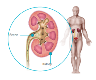


Ureteral Stent in kidney

Regular side effects of your stent 6-8

  • Pain, discomfort due to pressure that may increase when moving or after passing urine
  • Encrustation of the stent: the formation of a thin coating of stone material on the stent
  • Increased risk of a Urinary Tract Infection (UTI)
  • Bladder irritation symptoms include:

– Frequency

– Urgency

– Dysuria: painful or difficult urination

– Leakage

– Feeling of being unable to completely empty your bladder

  • Hematuria: having blood in your urine

Don’t forget! You have a foreign body in you that will have to be removed in the weeks following its insertion.

Potential stent complications 7,8

  • Migration or dislodgement of the stent
  • Obstruction of the stent
  • Rupture of the stent
  • Erosion of the mucosa

How long will my stent stay in? 5,9

JJ Stent are intended to be placed in the body temporarily. During the treatment process of urinary stones, the JJ stents are left in place from a few days to few months, depending on the case, after which they must be removed. The JJ stent will be removed remotely, through the urinary tract, usually under local anaesthetic at a consultation (few minutes intervention) or sometimes under general anaesthetic.

Living with a stent - work
Work:
If your job requires a significant amount of physical movement you may experience some pain and discomfort in your stomach and back area.
Living with a stent - sexual activity
Sex:
No specific contra-indications but some discomfort may occur during sexual activity. If your stent has a string, you may experience more discomfort and the stent may become dislodged.
 Living with a stent - Diet Diet:
Nutritional guidelines could be proposed according to the nature of the stone. Your diet can remain the same, but ensure that you are drinking plenty of fluids (2 liters per day) after the procedure. If you are taking any medications, ask your doctor if it is okay to consume alcoholic beverages.
 Living with a stent - having a social life Social Life:
Your social life should not be disrupted, but you may notice an increased frequency and urgency to use the restroom.
 Living with a stent - exercice Exercise:
Activity may cause pain and discomfort in your stomach area. Avoid vigorous exercise.
 Living with a stent - travelling Travel:
Before traveling, consult your doctor regarding your condition to see if it is safe. Your stent should not affect your ability to travel.
Close
JJ stent removal

JJ stent removal

How will my stent be removed? Learn more about quality of life with a JJ stent

References

1: Doersch KM, Elmekresh A, Machen GL, El Tayeb MM. The use of a string with a stent for self-removal following ureteroscopy: A safe practice to remain. Arab J Urol. 2018 Dec; 16(4): 435–440.
2: British Association of Urological Surgeon (BAUS). Telescopic insertion of removal of a stent from the ureter. 2017 https://www.baus.org.uk/_userfiles/pages/files/Patients/Leaflets/Ureteric%20stent%20insertion.pdf
3: NHS Hull University Teaching Hospitals - Ureteric Stent Removal under Local Anaesthetic –Discharge Advice. HEY-654/2018 https://www.hey.nhs.uk/patient-leaflet/ureteric-stent-removal-local-anaesthetic-discharge-ad- vice/
4: Baston EL, Wellum S, Bredow Z, Storey G, Palmer A, van Dellen D, Grey BR. Office-based ureteric stent removal is achievable, improves clinical flexibility and quality of care, whilst also keeping surgeons close to their patients. Cent European J Urol.2018;71(2):196-201. 
5: Isiris IFU
6: Collie J, Shah SS, Al-Hayek S, Durrant J, Saeb-Parsy K, Wiseman O. Introduction of a nurse led stent removal service using the single use isiris in a tertiary referral stone unit and the effect on patient waiting times. The Journal Of Urology. Vol. 201. 2019.

Your stent will be removed one of two ways:

If your stent has a withdrawal thread, you doctor will gently pull on it to remove the stent.1

If there is no withdrawal thread, the doctor will use an instrument called a cystoscope to grasp the stent coil located in your bladder and gently pull it through the urethra. 

You may experience some discomfort during the removal procedure and potentially experience slight pain few days after the stent has been removed.2,3

Isiris is an innovative single use stent removal solution with an integrated grasper, offering complete flexibility anywhere, anytime.4,5

Isiris demonstrated reduction in delays of stent removal and consequently significant quality of life improvements for patients.6

Discover more about Isiris α

Close
Metabolic assessment for stone disease

Metabolic assessment

Why does my surgeon talk about a “metabolic assessment”? Learn more about metabolic assessment

References

1: Can I help prevent kidney stones by changing what I eat or drink? 2017 https://www.niddk.nih.gov/health-information/urologic-diseases/kidney-stones/eating-diet-nutrition
2: Alelign T, Petros B. Kidney Stone Disease: An Update on Current Concepts. Adv Urol . 2018 Feb4;2018:3068365 

Why does my surgeon talk about a “metabolic assessment”?

The formation of urinary calculi is a condition that can develop over time and recur. Bad alimentary habits represent a major cause of the stones creation. Some dietetic measures related to drinks and diet can be taken to limit the recurrence.1

Following successful treatment of urinary stones, it is important to identify and manage the risk factors associated in the cause of urinary stone formation. This is called “metabolic assessment“.

The risk of recurrence is high. If patients do not apply prevention measures, the relapse rate of secondary stone formations is estimated to be 10–23% per year, 50% in 5–10 years, and 75% in 20 years after the first event in the patient.2

Based on dietary habits, underlying diseases, analysis of the removed stones and blood/ urinary tests, the urologist will be able to identify the causes of his patient’s urinary stone disease and put corrective measures into place. A medical drug can be prescribed but, in most cases, the treatment will be limited to diet recommendations. Dietary guidelines will be established by the doctor who can hand out a personalised explanatory sheet.

Sometimes it will be necessary to add to these first line exploratory examinations with more thorough exploration in a specialised environment.

Finally, urinary stone patients must perform regular check-ups of their kidneys and urinary tract, even in the absence of pain, e.g. by having an ultrasound examination every year, in case of any possible recurrence.

What does the “metabolic assessment” consist of?

  • Analysing the stone

This is carried out on the entire stone or fragments collected by the patient or urologist. The fragments must be stored in a container in a dry place and will be analysed using “Infrared Spectrophotometry”. If no stone or fragment has been recovered, a study will be performed of the nature of the urine crystals by examining the fresh urine, known as “Crystalluria”.

  • Dietary survey

This provides information about a urinary stone patient’s eating and drinking habits. The urologist or dietary specialist will enquire in particular about the quantity and nature of the drinks, but also about the number and nature of dairy products, the daily amount of animal protein (meat, fish and seafood, eggs, processed pork products), the amount of salt and consumption of certain foods that are especially high in oxalate (dark chocolate and cocoa, tea, walnuts and hazelnuts, spinach, jelly candies, etc.).

  • A fasting blood sample and a collection of urine over 24 hours.
    These biological examinations can be carried out in any analytical laboratory.
Close
Dietary guidelines for stone disease

Dietary guidelines

Learn about the recommendations that could reduce the risk. Learn more about dietary guidelines

References

1: D'Alessandro C, Ferraro PM, Cianchi C, Barsotti M, Gambaro G, Cupisti A. Which Diet for Calcium Stone Patients: A Real-World Approach to Preventive Care? Nutrients. 2019 May 27;11(5):1182. 

Drinks1

It is important to drink water in sufficient quantities. This dilutes your urine and reduces the risk of stones. If you are drinking enough water, you shall expect to produce approximately 2 Liters of clear or light yellow colored urine per day.

How much should you drink?

  • 2 litres per day, or more if it is a warm day or you undertake a sports activity.

When should you drink?

  • Every day, taking a drink regularly throughout the day.
  • Including at night before going to bed.
  • And during the night if you wake up.

What should you drink?

  • Liquids like tap or bottled water, coffee, herbal tea, etc. are allowed.
  • The quantity of fluid intake is more important than the quality
  • Two glasses of freshly squeezed orange juice is recommended.

Bad dietary habits are a major cause of calcium, oxalate and uric stones. Dietary management involves both drinks and food.

Food1

It is not a question of going on a diet but of adjusting your dietary habits. Too much calcium, salt, sugar, animal protein, oxalate and uric acid can increase the risk of stones.

Calcium intake

  • Calcium comes from water and dairy products.
  • Do not avoid milk, yogurt, fresh cheeses but you should aim for a balance - neither too much nor too little calcium.
  • Your intake should be anything from 800mg to 1 gram per day.
  • It is recommended that you have 2 to 3 portions of dairy products per day depending on the calcium content of your water (see table).
  • 1 glass of milk (15cl) = 1 yoghurt = 100g of cream cheese.

In real terms, calculate and then choose accordingly

  • If you drink 2 litres of water that is low in calcium (less than 20mg/l), you should consume around 800mg of calcium in the form of dairy products. 
  • If you drink 2 litres of water that is high in calcium (more than 400mg/l), you should limit your dairy intake.

Salt intake

  • Too much salt in your food will increase natriuresis (the amount of salt in your urine) which can increase the amount of calcium excreted in your urine.
  • You should limit your intake of foods and meals that are too salty (cured meats, fast food, ready meals).
  • Never add salt at mealtimes.

Uric acid intake

  • You may be prescribed to limit your intake of foods that contain uric acid: sugar (main source of uric acid), cured meats, offal (sweetbreads, kidneys, brain, liver, etc.), certain types of fish (herring, tuna, troutsardines in oil, anchovies, etc.) and seafood.
  • Eat fruit and vegetables regularly.

Animal protein intake

  • Animal protein is found in meat, fish, cured meats, poultry and also in eggs and seafood. 100g of meat corresponds to 100g of fish.
  • Limit animal protein intake and prefer proteins from vegetable sources.

Oxalate intake

  • Foods that are high in oxalate should be eaten in moderation, particularly chocolate and cocoa.
  • Also peanuts, walnuts, hazelnuts, almonds, asparagus, beetroot, rhubarb, spinach, sorrel, tea, figs.
  • Vitamin C in large quantities (500mg to 1g tablets) is not advised.
Close
Close

Sample request

Thank you

Your sample request has been received successfully

View desktop version